OPTICAL COHERENCE TOMOGRAPHY (OCT)
Optical Coherence Tomography (OCT) is an imaging technique used to provide unprecedented high-resolution and cross-sectional images of the eye. The OCT scan allows microstructures of the eye to be imaged and shows different colour-coded layers of the retina. It is particularly useful in the diagnosis and management of age-related macular degeneration, diabetic macular oedema, macular hole, and epiretinal membrane. Additionally, it is useful for central serous retinopathy and vitreo-macular traction syndrome. An OCT scan is also helpful for glaucoma. It has become a gold standard in monitoring the efficacy of intravitreal anti-VEGF injections (Eylea or Eylea HD) for age-related macular degeneration and tailoring treatment regimes.
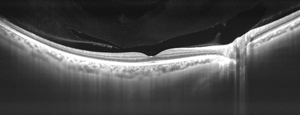
Ultrawide-field, high-resolution OCT scanning is available at City Eye Centre using the Plex Elite OCT (Carl Zeiss). It images all layers of the retina and choroid. The ultrawide-field imaging is particularly useful in myopia, retinal masses, and choroidal tumours. An OCT scan is very quick to perform and it is non-invasive and painless. Results are available instantaneously. It is an effective way for patients to gain a better understanding of their eye condition.
